How Do I Know If My Pineal Gland Is Damaged? Signs & Causes

You usually can’t tell if your pineal gland is damaged from symptoms alone. Most pineal gland changes, like calcification and small cysts, cause no symptoms at all and are found by accident on brain scans. The numbers show just how common these findings are.
A 2023 meta-analysis found pineal gland calcification in 61.65% of people studied, and a high-resolution MRI study found pineal cysts in 23% of healthy adults.
Real pineal problems that cause symptoms, such as large cysts or tumors, are much rarer. Pineal region tumors make up less than 1% of adult brain tumors and 3% to 8% of childhood brain tumors.
When they do cause symptoms, the warning signs tend to be specific: headaches, trouble moving the eyes up or down, double vision, nausea, and signs of fluid buildup in the brain.
This article explains what the pineal gland does, where it’s located, how to tell the difference between harmless findings and real problems, and when to see a doctor.
What Is The Pineal Gland?
The pineal gland is a tiny, pinecone-shaped gland in the brain. Its main job is producing melatonin, the hormone that helps control your sleep-wake cycle.
According to a Johns Hopkins radiology case review, the pineal gland normally has a pinecone-like shape and measures about 8 to 10 mm, roughly the size of a pea.
Where Is The Pineal Gland Located?
The pineal gland sits deep in the center of the brain, between the two halves (hemispheres).
It’s tucked near the back of a fluid-filled space called the third ventricle and close to the brainstem.
| Pineal Gland Fact | Detail |
|---|---|
| Location | Deep in the center of the brain, between the hemispheres |
| Size | About 8 to 10 mm, roughly pea-sized |
| Shape | Like a small pinecone |
| Main hormone | Melatonin |
| Main function | Helps regulate sleep and circadian rhythm |
This central location matters. Because the pineal gland sits right next to important pathways for brain fluid and eye movement, a large cyst or tumor there can press on nearby structures and cause specific symptoms.
What Is The Function Of The Pineal Gland?
The pineal gland’s best-understood role is controlling melatonin based on light and darkness.
Researchers note that about 80% of the gland is made up of cells whose main product is melatonin, which plays a major role in circadian rhythm.
Pineal gland hormones and their roles:
- Melatonin: rises in the evening and at night to help you sleep, and drops during the day.
- Hormone timing signals: in children, pineal region problems can sometimes affect hormones and puberty timing, which is one reason doctors watch for hormonal changes with pineal tumors.
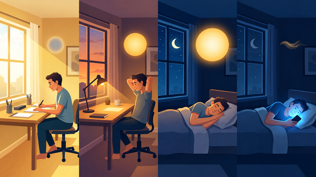
Light is the main signal the pineal gland responds to. Harvard Health Publishing explains that light exposure suppresses melatonin secretion, and blue light at night does so more powerfully.
What Is The Pineal Gland Third Eye?
The “third eye” idea comes from spiritual and philosophical traditions, not medicine.
Because the pineal gland responds to light and sits in the middle of the brain, some belief systems link it to intuition, spiritual awareness, or a “sixth sense.”
These ideas are popular online, but there’s no medical evidence linking pineal gland health to spiritual abilities.
Some supplements use this “third eye” idea in their marketing, and our in-depth Pineal Guardian X analysis looks at what’s actually behind those claims.
When we talk about pineal gland damage in this guide, we mean physical changes doctors can see on scans.
What Can Go Wrong With The Pineal Gland?
There are three main types of pineal gland findings people ask about:
| Pineal Finding | How Common | Usually Causes Symptoms? |
|---|---|---|
| Pineal gland calcification | Very common (about 61.65% in pooled research) | No |
| Pineal cyst | Common (up to 23% on high-resolution MRI) | Usually no |
| Pineal region tumor | Rare (under 1% of adult brain tumors) | Often yes, as it grows |
Let’s look at each one.
What Is A Calcified Pineal Gland?
A calcified pineal gland has tiny mineral deposits, mostly calcium and phosphorus, inside it. A 2023 systematic review describes these deposits as corpora arenacea.
How Common Is Pineal Gland Calcification?
Very common. The 2023 meta-analysis found a pooled prevalence of 61.65%. Calcification increases with age and is more common in men.
A 2023 Iranian CT study also linked smoking with higher odds of calcification.
What Are The Symptoms Of A Calcified Pineal Gland?
In most cases, there are none. Pineal calcification is usually found by accident on CT scans done for other reasons.
Researchers are studying possible links between calcification and:
- Lower melatonin production: a histology review suggests age-related changes, including calcification, may reduce melatonin synthesis.
- Alzheimer’s disease: some researchers have proposed calcification may play a role, though this remains under study.
But the evidence is mixed. Some researchers report melatonin levels aren’t affected by calcification at all.
About online “calcified pineal gland symptoms”: many websites list brain fog, poor sleep, low motivation, anxiety, or “blocked intuition.”
These symptoms have many possible causes, and there’s no good evidence that pineal calcification causes them in most people.
Can You Decalcify Your Pineal Gland?
There’s no scientific evidence that any method can decalcify the pineal gland.
We found no published human studies showing that diet changes, fluoride avoidance, supplements, or detox protocols reverse pineal calcification.
A 2001 study found fluoride accumulates in calcified pineal tissue of elderly people, but fact-checkers note there’s no clinical evidence fluoride harms the human pineal gland.
What you can do instead: support healthy melatonin by limiting bright and blue light at night, getting morning daylight, keeping a regular sleep schedule, and not smoking. To see what’s realistic, read our article on how to repair the pineal gland naturally.
What Is A Pineal Cyst?
A pineal cyst is a small, fluid-filled sac in the pineal gland. It’s usually benign (not cancer) and very common.
How Common Are Pineal Cysts?
It depends on how closely you look:
| Study Type | Prevalence |
|---|---|
| High-resolution MRI of healthy adults | 23% |
| Routine MRI in patients with neurological symptoms | 1.3% to 4.3% |
| MRI of asymptomatic healthy volunteers | Up to 10.8% |
| Autopsy studies | Up to 40% |
Most pineal cysts are tiny. One neurosurgery reference notes that more than 80% of pineal cysts are smaller than 10 mm and very rarely grow on follow-up imaging.
What Are Pineal Cyst Symptoms?
Most pineal cysts cause no symptoms at all. A clinical overview states that pineal cysts usually have no clinical implications and remain symptom-free for years.
When a pineal cyst does cause symptoms, it’s usually because it’s large enough to press on nearby structures. Reported symptoms include:
- Headache
- Dizziness or vertigo
- Double vision or blurred vision
- Trouble moving the eyes, especially looking up (Parinaud syndrome)
- Nausea and vomiting
- Hydrocephalus (fluid buildup in the brain)
The AJNR study explains that a cyst can occasionally enlarge the gland and compress nearby structures, including the passage that drains brain fluid, which can cause hydrocephalus and symptoms.
Do Pineal Cysts Cause Headaches?
This is a tricky question. Headache is the most common reason people get a brain MRI in the first place, so many pineal cysts are found in people who have headaches.
But that doesn’t mean the cyst caused the headache. In a UK study of 281 patients with pineal cysts, headache was the main reason for the MRI in 50.2% of cases, yet doctors didn’t find any symptoms attributable to the pineal cysts.
A study of children with pineal cysts also noted that, apart from rare symptoms like Parinaud syndrome and hydrocephalus, linking symptoms to the cyst is extremely difficult to prove.
Should I Be Worried About A Pineal Cyst?
Usually not. Here’s what the research shows:
- In the UK study, only 6% of pineal cysts changed size during follow-up, and changes were tiny (median of about 2 mm). No patients developed complications.
- The researchers concluded that incidental pineal cysts typically follow a benign course and recommended a single follow-up MRI at 12 months for adults without symptoms.
- A Turkish study of patients with pineal cysts found no significant size changes on follow-up scans.
When a pineal cyst may need more attention:
- It causes eye movement problems or double vision
- It causes hydrocephalus
- It’s growing on follow-up scans
- Your neurological symptoms are getting worse
- It has unusual features on imaging
One reference notes adults have about a 4% risk of cyst growth at 6 months, while children have about an 11% risk at 10 months, so children may need closer monitoring.
What Is Pineal Apoplexy?
Rarely, a pineal cyst can bleed internally and suddenly enlarge. This is called pineal apoplexy.
A neuroimaging reference explains that sudden bleeding can cause rapid onset of headache, vision problems, and hydrocephalus.
The AJNR study notes rare cases of sudden death from bleeding into a cyst.
This is very rare, but sudden, severe headache with vision changes or vomiting is an emergency.
What Is The Treatment For A Pineal Cyst?
Most pineal cysts need no treatment beyond, in some cases, a follow-up scan.
A clinical overview reports agreement that surgery should be considered for patients with hydrocephalus, worsening neurological symptoms, or cyst enlargement.
| Situation | Typical Approach |
|---|---|
| Small cyst, no symptoms | No treatment; possibly one follow-up MRI |
| Stable cyst over time | Usually discharged from follow-up |
| Child with a cyst | Often closer monitoring |
| Cyst causing hydrocephalus | Surgery may be considered |
| Cyst with worsening symptoms or growth | Surgery may be considered |
What Is A Pineal Gland Tumor?
A pineal tumor is an abnormal growth in or around the pineal gland. Unlike cysts, tumors can grow and cause problems by pressing on nearby brain structures.
How Common Are Pineal Tumors?
Pineal region tumors are rare. Columbia University Neurosurgery reports they make up less than 1% of adult brain tumors and 3% to 8% of childhood brain tumors.
In children, they tend to appear between ages 10 and 20, and in adults, after age 30.
UCSF’s Brain Tumor Center reports they make up 2.6% of primary brain tumors in children aged 0 to 14, and 1.2% in teens and young adults aged 15 to 39.
What Types Of Pineal Tumors Are There?
| Tumor Type | What It Is |
|---|---|
| Germ cell tumors (including germinomas) | The most common pineal region tumors; can be benign or malignant |
| Pineocytoma | Usually slow-growing and less aggressive |
| Pineoblastoma | Fast-growing and aggressive, seen most often in young children |
| Pineal tumor of intermediate differentiation | Between pineocytoma and pineoblastoma |
| Gliomas | Arise from nearby glial cells; range from low to high grade |
What Are Pineal Gland Tumor Symptoms?
Stony Brook Medicine lists these possible symptoms of pineal region tumors:
- Headaches
- Blurred or double vision
- Trouble moving the eyes up or down
- Hydrocephalus
- Nausea and vomiting
- Memory or thinking changes
- Sleep problems
- Balance or coordination issues
- Hormonal changes
Symptoms often develop gradually, from weeks to years.
How Are Pineal Tumors Treated?
Treatment depends on the tumor type. Columbia Neurosurgery notes that surgery is the standard first step for most pineal region tumors.
Other treatments may include:
- Radiation therapy: some germ cell tumors respond very well to radiation.
- Chemotherapy: may be effective for germ cell tumors and some other types.
- Shunts or other procedures: to relieve hydrocephalus.
UCSF notes that if a germinoma is suspected, a biopsy is usually needed to confirm the diagnosis before treatment.
Outlook varies widely. For pineocytomas, a Johns Hopkins case review reports an excellent 5-year survival rate of 85% to 100% after complete surgical removal.
Pineal Cyst Vs Tumor: What’s The Difference?
| Feature | Pineal Cyst | Pineal Tumor |
|---|---|---|
| What it is | Fluid-filled sac | Abnormal tissue growth |
| How common | Very common | Rare |
| Usually benign? | Yes | Can be benign or malignant |
| Usually grows? | Rarely | Can grow over time |
| Usually causes symptoms? | No | Often, as it grows |
| Typical treatment | Observation or none | Surgery, radiation, chemotherapy |
| Diagnosis | MRI | MRI, sometimes biopsy |
Why it can be confusing: a study of children with pineal cysts notes that telling a benign cyst apart from a tumor can be challenging even with advanced MRI.
That’s why doctors sometimes recommend contrast-enhanced MRI or follow-up scans to be sure.
How Do Doctors Check For Pineal Gland Damage?
You can’t diagnose pineal gland problems at home. Doctors use:
| Test | What It Shows |
|---|---|
| Neurological exam | Eye movements, balance, reflexes, and other signs |
| CT scan | Clearly shows calcification |
| MRI | Best for cysts and tumors |
| MRI with contrast | Helps tell cysts from tumors |
| Eye exam | Checks for swelling of the optic nerve from pressure |
| Blood tests | Tumor markers for some germ cell tumors |
| Biopsy | Confirms tumor type when needed |
What Is Pineal Gland Dysfunction?
“Pineal gland dysfunction” isn’t a common formal diagnosis. People often use the term to mean problems with melatonin and sleep.
Real causes of disrupted melatonin and sleep rhythms are usually not pineal damage. They include:
- Bright or blue light at night
- Irregular sleep schedules or shift work
- Jet lag
- Stress and anxiety
- Certain medications
- Sleep disorders
Pineal region tumors can cause sleep problems and hormonal changes, but they’re rare. If you have ongoing sleep problems, a doctor can help find the cause. And if a racing mind is what keeps you up, these tips for calming your mind at night can help.
When Should You See A Doctor?
See a doctor if you have:
- Ongoing or worsening headaches
- Blurred or double vision
- Trouble looking up or down
- Problems with balance or coordination
- Memory or thinking changes
- Ongoing sleep problems
- Early or delayed puberty in a child
- A scan report mentioning a pineal finding you don’t understand
Get emergency care right away if you have:
- A sudden, severe headache
- Sudden vision changes or double vision
- Repeated vomiting with headache
- Confusion, extreme drowsiness, or fainting
- Seizures
- Weakness on one side of the body
These can be signs of serious problems, including hydrocephalus, bleeding, or stroke.
How Can You Support Pineal Gland Health?
You can’t prevent most pineal findings, but you can support healthy melatonin and sleep:
| Habit | Why It Helps |
|---|---|
| Limit screens 2 to 3 hours before bed | Harvard Health recommends this to protect melatonin |
| Use dim, warm light in the evening | Red light is less likely to suppress melatonin |
| Get morning daylight | Helps set your body clock |
| Keep a regular sleep schedule | Supports predictable melatonin release |
| Keep your bedroom dark | Even dim light can affect melatonin |
| Don’t smoke | Linked to higher pineal calcification |
Some people also consider a pineal gland support formula, but no supplement can replace these everyday habits.
What Is The Bottom Line On Pineal Gland Damage?
For most people, pineal gland findings are harmless. Calcification affects a majority of adults and usually causes no symptoms.
Pineal cysts are common, rarely grow, and seldom need treatment. Pineal tumors are rare, but they tend to cause specific warning signs as they grow, such as headaches, double vision, trouble moving the eyes up or down, nausea, and balance problems.
You can’t diagnose pineal damage from vague symptoms like brain fog or low motivation, and you can’t decalcify your pineal gland with supplements or detox methods.
If a scan mentions a pineal finding, ask your doctor what it means and whether follow-up is needed.
If you develop sudden severe headaches, vision changes, or vomiting, seek emergency care.
And to support your pineal gland’s main job, focus on healthy light exposure and consistent sleep habits.
What Do People Ask Most About Pineal Gland Damage?
How Do I Know If My Pineal Gland Is Damaged?
Pineal gland damage usually can’t be detected from symptoms alone and is typically found on brain scans. Calcification and small cysts usually cause no symptoms. Warning signs of larger problems include headaches, double vision, trouble looking up, nausea, and balance issues.
Where Is The Pineal Gland Located?
The pineal gland is located deep in the center of the brain, between the two hemispheres. It sits near the third ventricle and close to the brainstem. It’s about 8 to 10 mm in size.
What Is The Function Of The Pineal Gland?
The pineal gland’s main function is producing melatonin, which helps regulate sleep and circadian rhythm. Melatonin rises at night and drops during the day. Light exposure, especially blue light, suppresses melatonin.
What Are Pineal Cyst Symptoms?
Pineal cysts usually cause no symptoms. Large cysts can occasionally cause headaches, dizziness, double vision, trouble moving the eyes upward, nausea, and hydrocephalus. Linking headaches to small cysts is difficult to prove.
Should I Be Worried About A Pineal Cyst?
Pineal cysts are usually nothing to worry about. In a UK study of 281 patients, only 6% changed size and none developed complications. Researchers recommended a single follow-up MRI at 12 months for adults without symptoms.
What Are Pineal Gland Tumor Symptoms?
Pineal gland tumor symptoms may include headaches, blurred or double vision, trouble moving the eyes, nausea, vomiting, balance problems, memory changes, sleep problems, and hormonal changes. These tumors are rare, making up under 1% of adult brain tumors.
What Is The Difference Between A Pineal Cyst And A Tumor?
A pineal cyst is a common, usually harmless fluid-filled sac, while a pineal tumor is a rare abnormal growth that can be benign or malignant. Cysts rarely grow, while tumors can enlarge and cause symptoms. MRI helps tell them apart.
What Are The Symptoms Of A Calcified Pineal Gland?
A calcified pineal gland usually causes no symptoms and is very common, affecting about 61.65% of people in pooled research. Researchers are studying possible links with melatonin and Alzheimer’s, but online symptom lists aren’t backed by strong evidence.
Is The Pineal Gland The Third Eye?
The pineal gland third eye idea comes from spiritual traditions, not medicine. The gland responds to light and produces melatonin, but there’s no medical evidence linking it to intuition or spiritual abilities.
References
- Belay DG, Worku MG. Prevalence of Pineal Gland Calcification: Systematic Review and Meta-Analysis. Systematic Reviews. 2023;12:32.
Reports a pooled pineal gland calcification prevalence of 61.65% and examines associations with age, sex, and ethnicity. - Arendt J, Aulinas A. Physiology of the Pineal Gland and Melatonin. Endotext. Updated October 30, 2022.
Reviews pineal gland anatomy, melatonin production, circadian rhythms, light-dark signaling, and factors that influence melatonin secretion. - Jalali Z, et al. Cross-sectional analysis of potential risk factors of the pineal gland calcification. BMC Endocrine Disorders. 2023.
Examines potential factors associated with pineal calcification, including age, sex, smoking, and metabolic factors. - Luke J. Fluoride deposition in the aged human pineal gland. Caries Research. 2001;35(2):125-128.
Studied fluoride and calcium concentrations in pineal glands from 11 elderly cadavers and found a positive correlation between pineal fluoride and calcium. The study does not establish that fluoride damages pineal function. - The natural history of pineal cysts in children and young adults. Journal of Neurosurgery: Pediatrics.
Follow-up of 106 patients younger than 25 found that most pineal cysts remained stable, while cyst growth or imaging changes were more common among younger children.